Basic Cell Poster Set
These are the five posters within the Basic Cell Set:
This Basic Cell Set of posters provides your students broad examples of what can be understood from light microscopy of cells. These micrographs were all taken at 400X magnification, and each image demonstrates different cells and cell components. This set is an excellent complement to the Dynamic Cell Models for classrooms short on microscopes for their students. The 5 posters are:
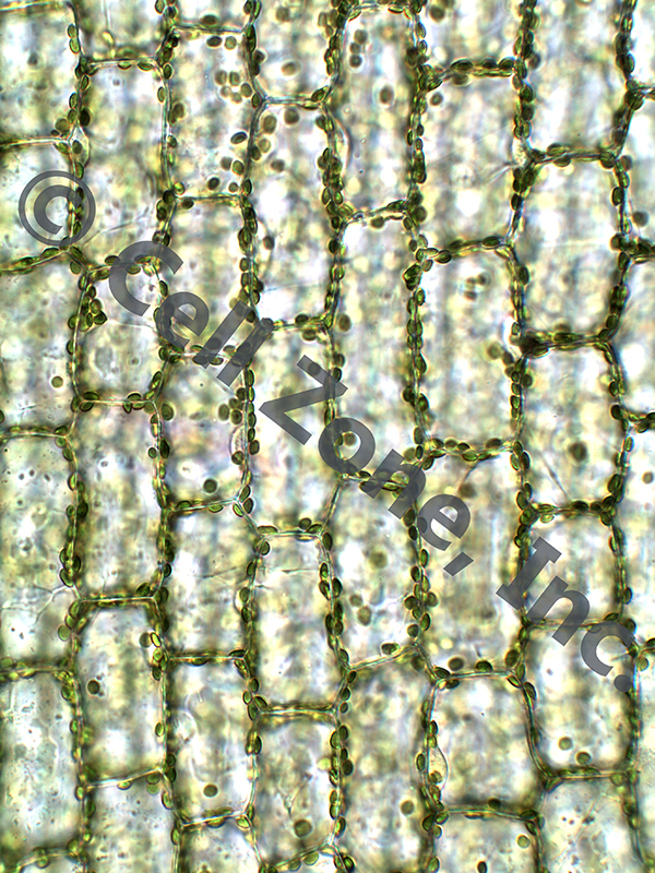
- Elodea canadensis: These plant cells can be used to see cell walls, cell membranes, chloroplasts, and cytosol. Some nuclei are also visible. The central vacuole is apparent when viewing live cells as the area avoided during cytoplasmic streaming. These cells are unstained.
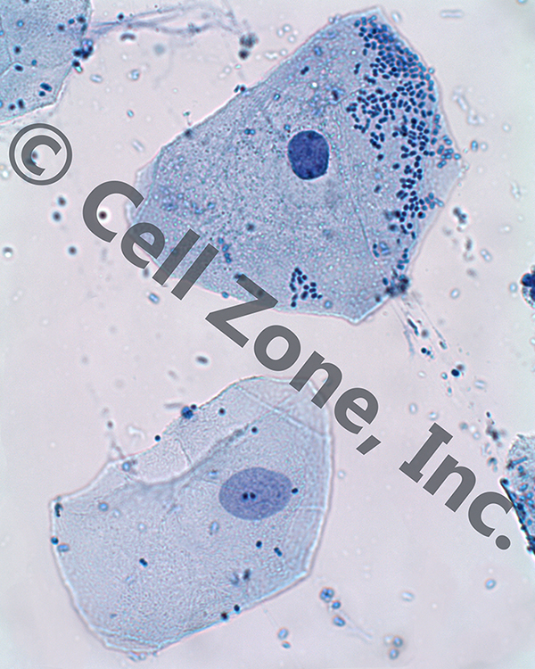
- cheek (with bacteria): These human (animal) cells can be used to view cytoplasm, nuclei, and cell membranes (only as the edge of the cytoplasm). These can also be used to show that animal cells lack cell walls. The methylene blue stain used on these cells also reveals bacillus and coccus bacteria stuck to the cheek cells and slide.
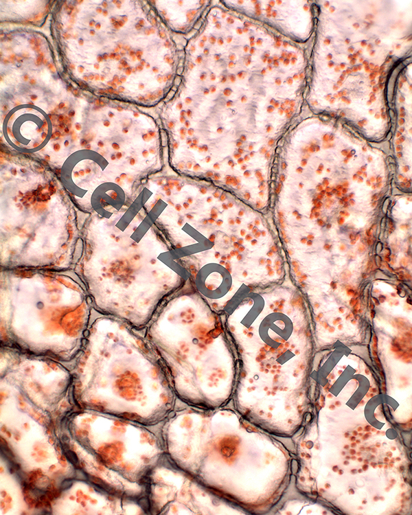
- red pepper: These plant cells are from the peel of a red pepper, which is a very tough portion of the fruit. Therefore, these cells demonstrate a particularly thick cell wall. Also visible are the red chromoplasts and plasmodesmata running through the thick cell walls. Some nuclei are also detectable, although these cells were not stained.
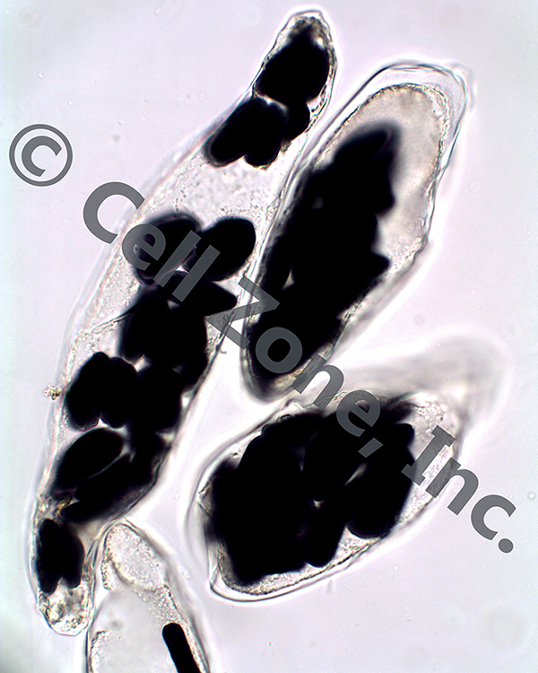
- banana cells: These plant cells were taken from the fruit of the banana. The cells were stained with iodine to reveal the starch contained within their amyloplasts. The cell walls (very thin), cell membrane and cytoplasm, and amyloplasts are visible. Note that we also have posted a free download of Dawn's Journal article in the American Biology Teacher on using banana cells to study carbohydrates on the "Resources" page. This article (and the associated classroom handout by Jon Darkow) are excellent companions to this poster in your classroom.
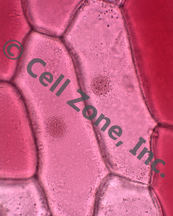
- red onion: These plant cells were taken from the thin purple layer of a red onion. They were coverslipped in iodine to add some contrast to their nuclei, but no starch is present. The cell walls, cytoplasm, and nuclei are visible.
Each poster pairs with a lesson plan to turn the poster into an active learning classroom tool.
Basic Cell Set, PST-cell
5 posters with 5 frames for $155 + shipping.
The lesson plans for these posters can be found on the Visual Microscopy Learning Kit Lesson Plan download page.
Now Reduced Prices!
When everything else costs more, we have lowered prices on our bestsellers to help your students learn.
Cell Zone, Inc.
21643 Deer Grass Drive
Escondido, CA 92029
Phone: +1 (413) 427-1214
Fax: +1 (413) 200-3250
E-mail: dawn@cellzone.org